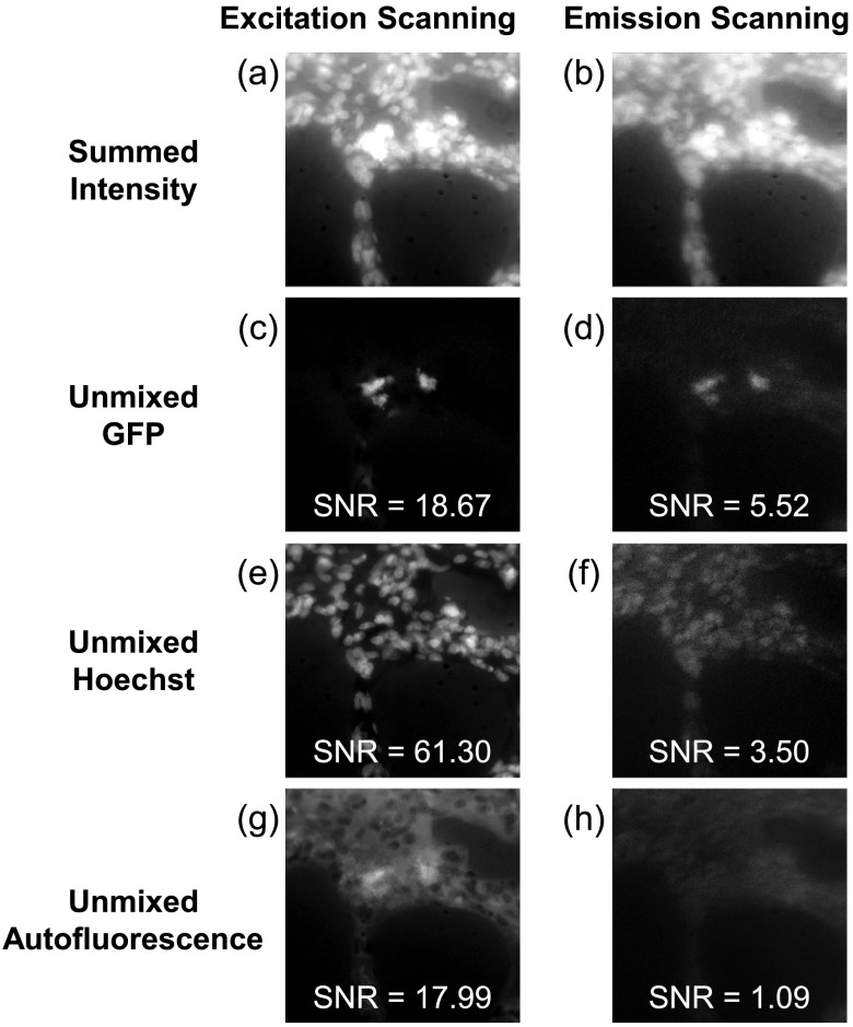
Fig. 5.
Linear unmixing of spectral image stacks allowed identification of GFP, Hoechst, and autofluorescence in both excitation- and emission-scanning hyperspectral image sets. The summed fluorescence intensity (a and b) was used to visualize raw image data and revealed improved clarity of nuclei and GFP in the excitation-scanning image compared with the emission-scanning image. The GFP emission was better localized using excitation scanning compared with emission scanning (c and d). In addition, nuclei were better resolved in the excitation-scanning image compared with the emission-scanning image (e and f). Finally, autofluorescence was more clearly delineated using excitation scanning than emission scanning (g and h). Excitation scanning provided a higher signal-to-noise ratio (SNR) for all three fluorophores measured. In specific, the Hoechst displayed a 20-fold increase in SNR with excitation scanning, compared with emission scanning. Additionally, GFP possessed a 3-fold increase in SNR with excitation scanning, compared with emission scanning.

