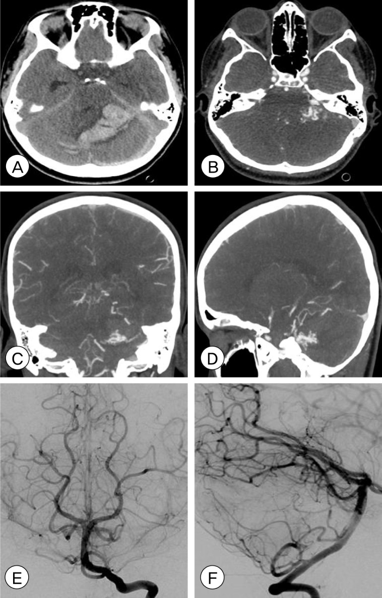
Fig. 1.

Pre-operative (A) non-contrast brain computed tomography (CT), axial view, shows a large cerebellar hematoma with severe effacement of the fourth ventricle, resultant hydrocephalus, and tonsillar herniation. Brain CT angiography (CTA), axial (B), coronal (C), and sagittal (D) views shows a Spetzler-Martin Grade II, supplementary grade I left cerebellar arteriovenous malformation (AVM) measuring 12×8 mm in size, supplied by the left superior cerebellar artery (SCA) and anterior inferior cerebellar artery (AICA) and draining into the left transverse and straight sinuses. Post-operative angiography with left vertebral artery injection, AP (E) and lateral (F) views, shows no residual AVM, consistent with complete microsurgical obliteration.
