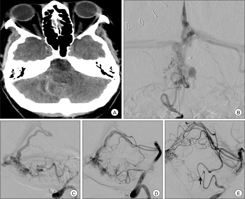
Fig. 2.
Pre-operative (A) non-contrast brain CT, axial view, shows diffuse cerebellar subarachnoid hemorrhage and intraventricular hemorrhage of the fourth ventricle resulting in obstructive hydrocephalus. Pre-operative angiography with right vertebral artery injection, AP (B) and lateral (C) views, and left vertebral artery injection, lateral view (D) shows a Spetzler-Martin Grade II, supplementary grade III right cerebellar arteriovenous malformation (AVM) measuring 14×15×13 mm in size, supplied by the bilateral superior cerebellar arteries (SCAs) and posterior inferior cerebellar arteries (PICAs) and draining into the vein of Galen. There are multiple feeding artery aneurysms on the right PICA as well as several venous dilatations of the deep draining vein. After pre-operative polyvinyl alcohol (PVA) particle embolization through the right PICA, the feeding artery was occluded with coils (arrow) as shown on this DSA right vertebral artery injection lateral view (E).

