
Fig. 2. Inter-relationship between Treg subsets.
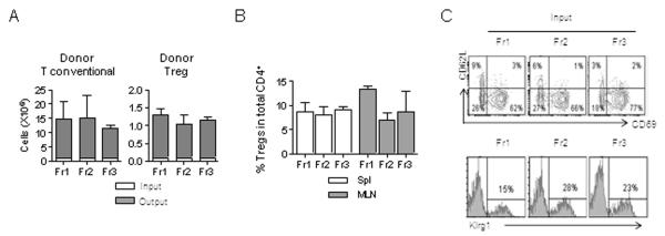
Adult TCRα−/− mice were adoptively transferred with a mixture (1:10) of RFP+ Tregs from purified subsets (Fr1-3, Klrg1−) and Treg-depleted conventional CD4+ T cells. The persistence and phenotype of the donor cells were assessed 3-4 weeks post-transfer. (A) Expansion of the donor T-conventional and Treg cells where the total numbers of injected (input) and recovered (output) cells from the spleen were enumerated. (B) Spleen and MLN were examined for donor Tregs as a fraction of the total CD4+ T-cell compartment. (C) The phenotype of donor Tregs from recipients that received the indicated fractions of Klrg1-depleted Fr1, Fr2, or Fr3 RFP+ donor Treg cells. Shown at the top is the expression of CD62L and CD69 and at the bottom is the expression of Klrg1 by donor Tregs.