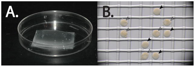
Figure 5.

Nitex injection plates. A. The Nitex mesh is affixed to the bottom of the petri dish with vacuum grease. The FSW is confined to a mound of water within the boundaries of the mesh. B. Sea star eggs (black arrowheads) and oocytes (white arrowheads) fit within the mesh. Note the lack of GVs in the eggs when compared to the oocytes.
