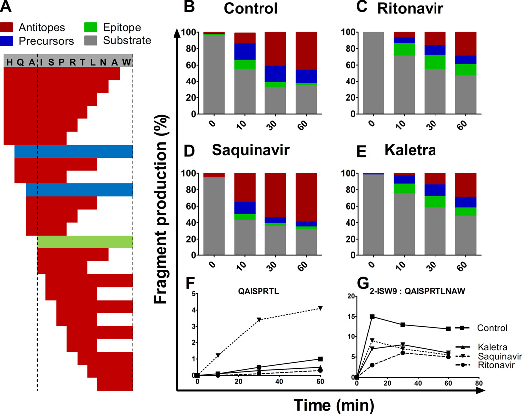
FIGURE 2.
HIV PIs alter peptide degradation patterns. 3ISW9 (HQAISPRTLNAW) peptide containing HLA-B57-restricted ISW9 epitope (ISPRTLNAW) was degraded in PBMC cytosolic extracts preincubated with DMSO (A, B) or 10 µM of Ritonavir (C), Saquinavir (D) or Kaletra (E). Resulting degradation products at 0, 10, 30 and 60 min were analyzed by LC-MS-MS. Degradation peptides were categorized as substrate 3-ISW9 (grey), epitope ISW9 (green), precursors -peptides that include the epitope (blue) or antitopes (peptides including only part of the epitope (red) (A–E). Each peptide identified by a specific mass and charge corresponds to a peak of a specific intensity and the proportion of each category of peptides to the total peak intensity (ranging from 7.1E+8 to 8.4E+8 at a given time point) was calculated at each time point in the presence of DMSO (B), Ritonavir (C), Saquinavir (D) or Kaletra (E). Percentage of antitope QAISPRTL (E) and precursor 2-ISW9 (QAISPRTLNAW) (F) production over time upon PI treatment. This figure is representative of one of three independent experiments using PBMC extracts from 3 different donors and run in duplicates on the mass spectrometer.

