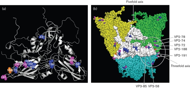
Fig. 1.
(a) O1K reduced structure (cartoon) showing position of critical amino acid residues of five neutralizing antigenic sites in blue. Residue VP2-188 shown in orange and VP2-78 shown in purple have been identified using bovine mAbs (Barnett et al., 1998). Additional residues mutated in this study are shown in magenta. (b) Three-dimensional structure (external surface) of the O1K (reduced) showing changes in antigenic sites 2 and 4 within the circle. Changes in amino acids forming the five neutralizing antigenic sites (5M) in the central protomer (shown in white) are shown in blue. Amino acids at position VP3-85 (close to VP3-58 that defines antigenic site 4), VP2-191 (located at the threefold axis) and VP2-74 (close to VP2-72 that defines antigenic site 2) are shown in magenta, VP2-188 in orange and VP2-78 in purple.

