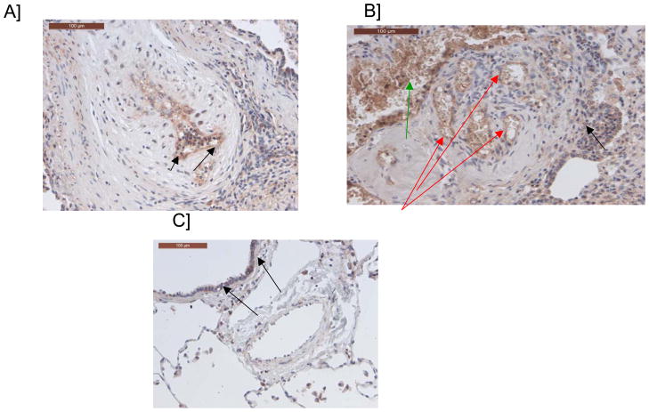
Figure 2. Immunostaining of pulmonary arteries with a cerebellin 2 antibody.
A/ Endothelial labeling of a pulmonary artery with intimal fibrosis (see arrows) B/ Endothelial labeling of a plexiform lesion (red arrows), labeling of intravascular cellular elements (erythrocytes, leukocytes) (green arrow), background-staining in some areas (black arrow); C/ Pulmonary artery from a control patient without labeling. Scattered bronchial cells are labeled (black arrows).

