
Figure 2.
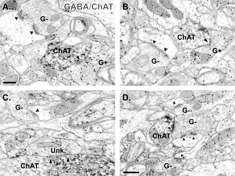
Electron micrographs of tissue double immunostained for GABA (immunogold) and ChAT (immunoperoxidase) in the monkey putamen. A,B: Photomicrographs demonstrating GABA-positive (G+) terminals forming symmetric synapses (arrows) with medium-sized ChAT-labeled dendrites. Note that a GABA-negative (G−) terminal in each micrograph is forming an asymmetric synapse (black arrowheads) with a spine. C: Example of a terminal categorized as “symmetric/unknown” (Unk.) that forms a symmetric synapse (black arrows) with a large-sized ChAT-positive dendrite. A GABA-negative (G−) terminal forming an axo-spinous synapse (arrowhead) is also visible. D: Example of a GABA-negative terminal forming an asymmetric synapse onto a small-sized ChAT-labeled dendrite and an unlabeled spine (arrowheads). Other GABA-negative (G−) terminals are also visible in this tissue. Scale bar = 0.2 μm in A (applies to A,B); 0.5 μm in D (applies to C,D).