
Figure 7.
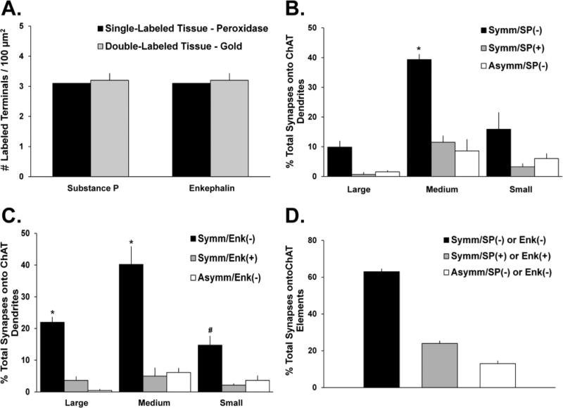
Proportion of total synaptic inputs onto ChIs from SP- and Enk-ir terminals. A: Comparison of the mean densities of SP- or Enk-positive terminals as revealed with the pre-embedding immunogold method in double-immunostained striatal tissue of three monkeys (SP/ChAT-left gray bar; Enk/ChAT-right gray bar) with the density of these terminals in striatal tissue from one monkey single labeled for each peptide with the immunoperoxidase method. The density of labeled terminals for either SP or Enk is the same irrespective of the marker used in single or double immunolabeling reactions. B,C: Mean percentages (± SEM) of total terminals in synaptic contact with ChI dendrites in the monkey putamen. Terminals are categorized based on their type of synaptic specializations (symmetric vs. asymmetric) and neuropeptide immunoreactivity (SP+ vs. SP− in B; Enk+ vs. Enk− in C). In B, *, P < 0.001 Symm/SP(−) versus Symm/SP(+) and Asymm/SP(−) on medium dendrites. In C, *, P < 0.001, Symm/Enk(−) versus Symm/Enk(+) and Asymm/Enk(−) on large or medium dendrites; #, Symm/Enk(−) versus Symm/Enk(+) on small dendrites. D: Mean percentages of synaptic inputs onto ChIs that originate from SP+ and Enk+ collaterals of GABAergic striatal projection neurons (Symm/SP+ or Enk+) or other sources of putatively GABAergic terminals (Symm/SP− and Enk−) and putatively glutamatergic boutons (Asymm/SP− and Enk−).