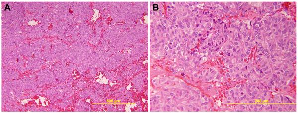
Fig. 2.

Routine H&E histologic evaluation of the adrenal gland shows a pheochromocytoma. The neoplasm has an organoid growth pattern with hemorrhage separating islands of tumor cells (A) [low-power, 100x]. The individual tumor cells have abundant eosinophilic cytoplasm (B) [high-power, 400x].
