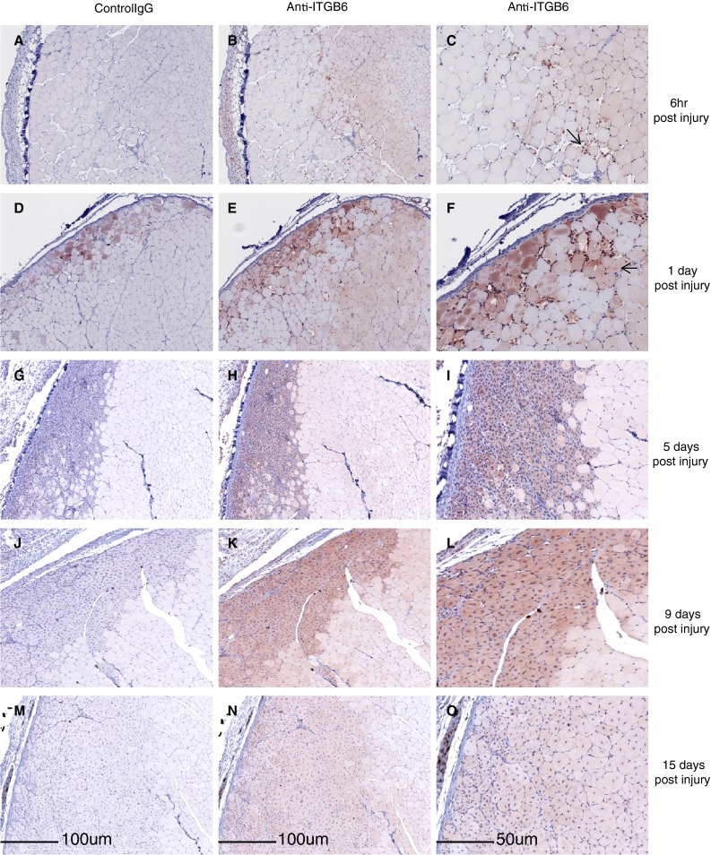
Fig. 4.

Mouse muscle post freeze injury time course. a 6 h post-injury control IgG. b 6 h post-injury. Light ITGB6 staining is detected in undamaged myocytes, c 6 h post-injury, magnified area of 4b showing intensely stained immune cells (arrow) d 1 day post-injury control IgG. e 1 day post-injury ITGB6 staining is detected in undamaged fibers. f. 1 day post-injury magnified area of 4e. ITGB6 staining is detected in some of the damaged fibers (arrow). g 5 days post-injury IgG control. h 5 days post-injury ITGB6 staining is detected in regenerating fibers (boxed region). i 5 days post-injury magnified area of 4h. j 9 days post-injury IgG control. k 9 days post-injury. Regenerated fibers still have centrally located nuclei and stain for ITGB6. (boxed region). l 9 days post-injury magnified area of 4k. m 15 days post-injury control IgG. n 15 days post-injury newly formed fibers with centrally located nuclei express low levels of ITGB6. o 15 days post-injury magnified area of 4n
