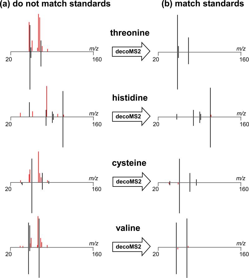
Figure 3. decoMS2 applied to amino acid standards.
Amino acid standards were mixed and analyzed by LC/MS2, using a 9 Da MS2 isolation window. Because the amino acid standards were inadequately separated, each of their MS2 spectra were contaminated by additional precursors in the collision cell. (a) The experimental MS2 spectra for 4 representative amino acids are shown on the top of each plot. The standard MS2 spectra for each of these amino acids as obtained from pure model compounds is shown on the bottom. Fragments that match in the 2 spectra are colored black, while fragments that do not match are colored red. (b) After the application of decoMS2, the top experimental MS2 spectra of each amino acid are highly consistent with the MS2 spectra from their respective amino acid standards.

