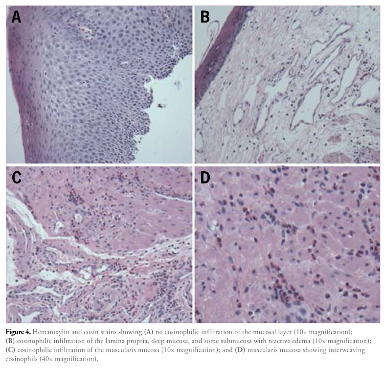
Figure 4.
Hematoxylin and eosin stains showing (A) no eosinophilic infiltration of the mucosal layer (10x magnification); eosinophilic infiltration of the lamina propria, deep mucosa, and some submucosa with reactive edema (10x magnification); eosinophilic infiltration of the muscularis mucosa (10x magnification); and (D) muscularis mucosa showing interweaving eosinophils (40x magnification).

