. 2013 Oct 7;42(24):9207–9218. doi: 10.1039/c3cs60198f

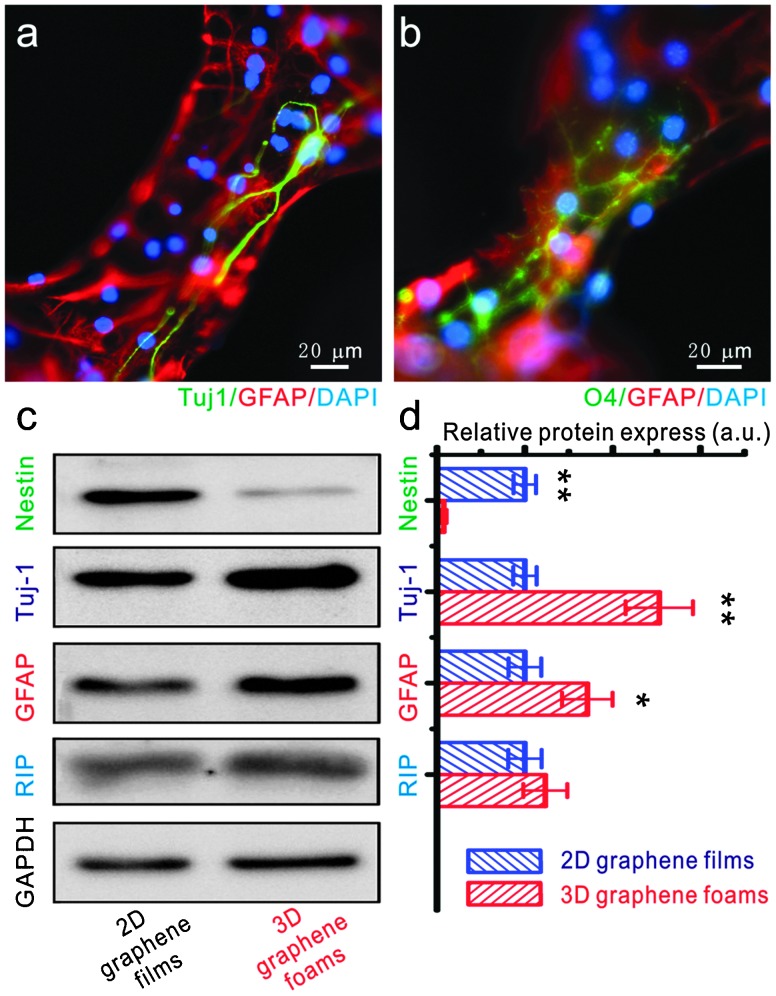
Fig. 3. The differentiation of NSCs on a 3D graphene foam scaffold. (a and b) Fluorescence images of differentiated NSCs that were immunostained for Tuj-1 (marker for neurons – green, a), GFAP (marker for astrocytes – red, a and b), O4 (marker for oligodendrocytes – green, b) and DAPI (marker for nuclei – blue, a and b). Western blot analysis of nestin (marker for NSCs), Tuj-1, GFAP and RIP (marker for oligodendrocyte) protein expression of differentiated NSCs on 2D graphene films and 3D graphene foam scaffold. (d) Relative optical densities of nestin, Tuj-1, GFAP and RIP bands shown in (c). Reproduced with permission from ref. 25 (copyright 2012, Nature Publishing Group).

Fig. 3