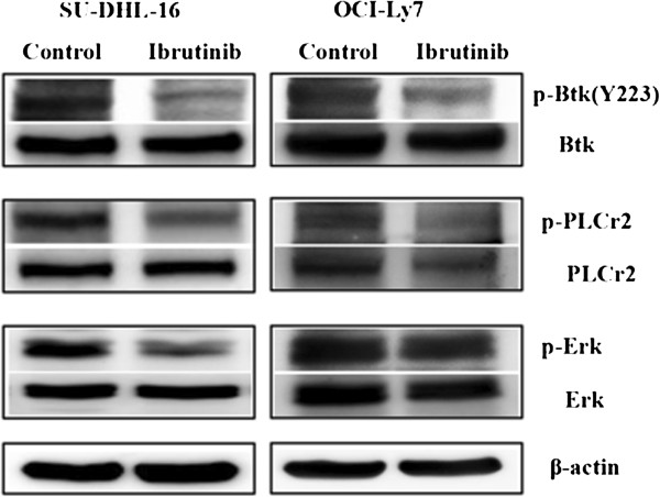
Figure 5.
Phosphorylation of ERK predicted the different response to ibrutinib. Cells (1 × 10⁵/ml) were incubate with ibrutinib at a concentration of 10 μM for 24 hours, then lysed using RIPA buffer for the total protein. The expression of p-Btk, p-PLCγ2, p-ERK and Btk, PLCγ2, ERK protein was detected by Western Blot. β-actin was included as a loading control. Results were shown from one of three experiments and the representative results were shown in the figures.

