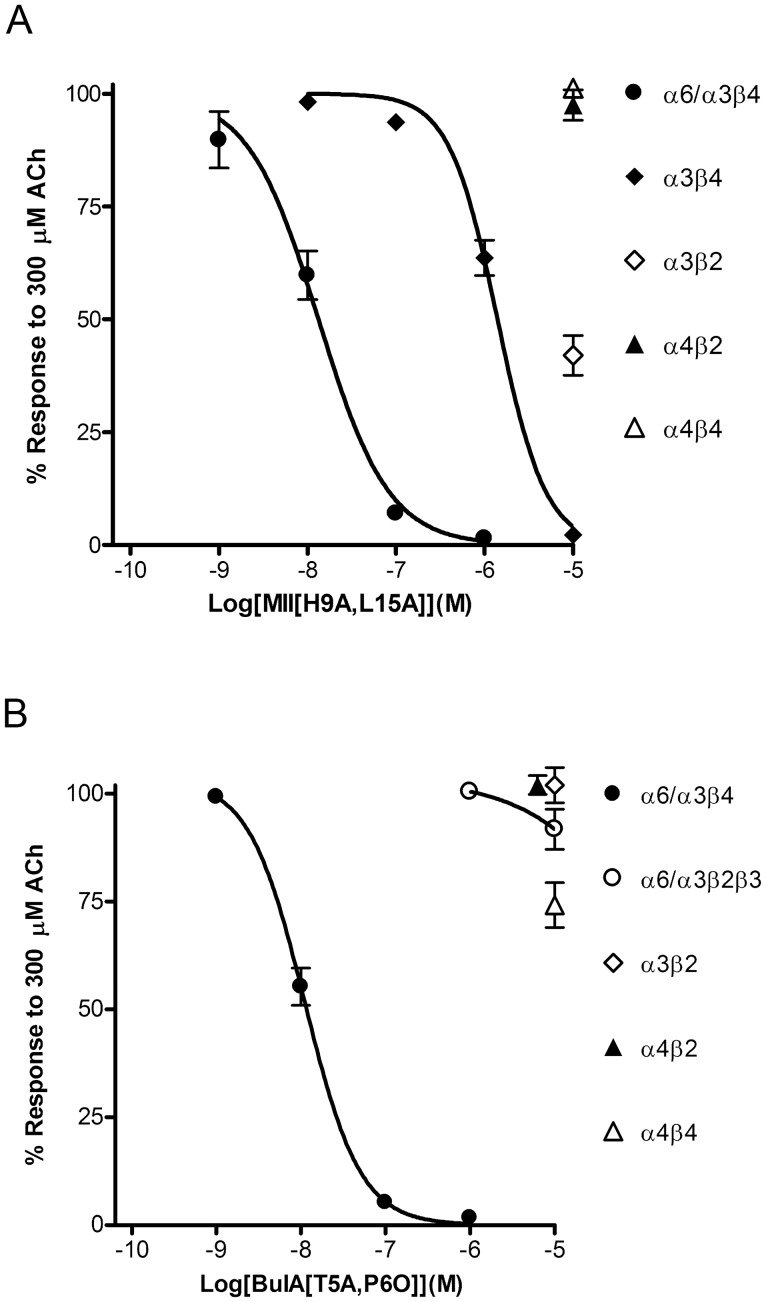
Figure 4. Concentration-response analysis of the inhibition of cloned human nAChRs expressed in X. laevis oocytes by α-Ctxs MII[H9A,L15A] and BuIA[T5A,P6O].
Oocytes expressing the indicated nAChRs were subjected to TEVC as described in “Materials and Methods” and the IC50 values for inhibition of the responses to ACh by each α-Ctx analog determined by fitting the data to the Hill equation. A, MII[H9A,L15A] inhibited α6/α3β4 with an IC50 value of 13.3 (9.7–18.1) nM (n = 4) and α3β4 with an IC50 value of 1.4 (1.1–1.7) μM (n = 4). Compared to controls, the responses after a 5 min exposure to 10 μM toxin were 42±4% (n = 7) for α3β2, 93±5% (n = 7) for α4β2, and 101±1% (n = 4) for α4β4 receptors. B, BuIA[T5A,P6O] inhibited α6/α3β4 with an IC50 value of 11.1 (9.1–13.6) nM and α6/α3β2β3 with an IC50 value >10 μM. The responses after a 5 min exposure to 10 μM toxin were 102±4% (n = 4) for α3β2, 102±2% (n = 4) for α4β2, and 74±5% (n = 5) for α4β4 receptors. For clarity, the symbols for inhibition of α3β2 and α4β2 receptors are shown staggered to avoid overlap.

