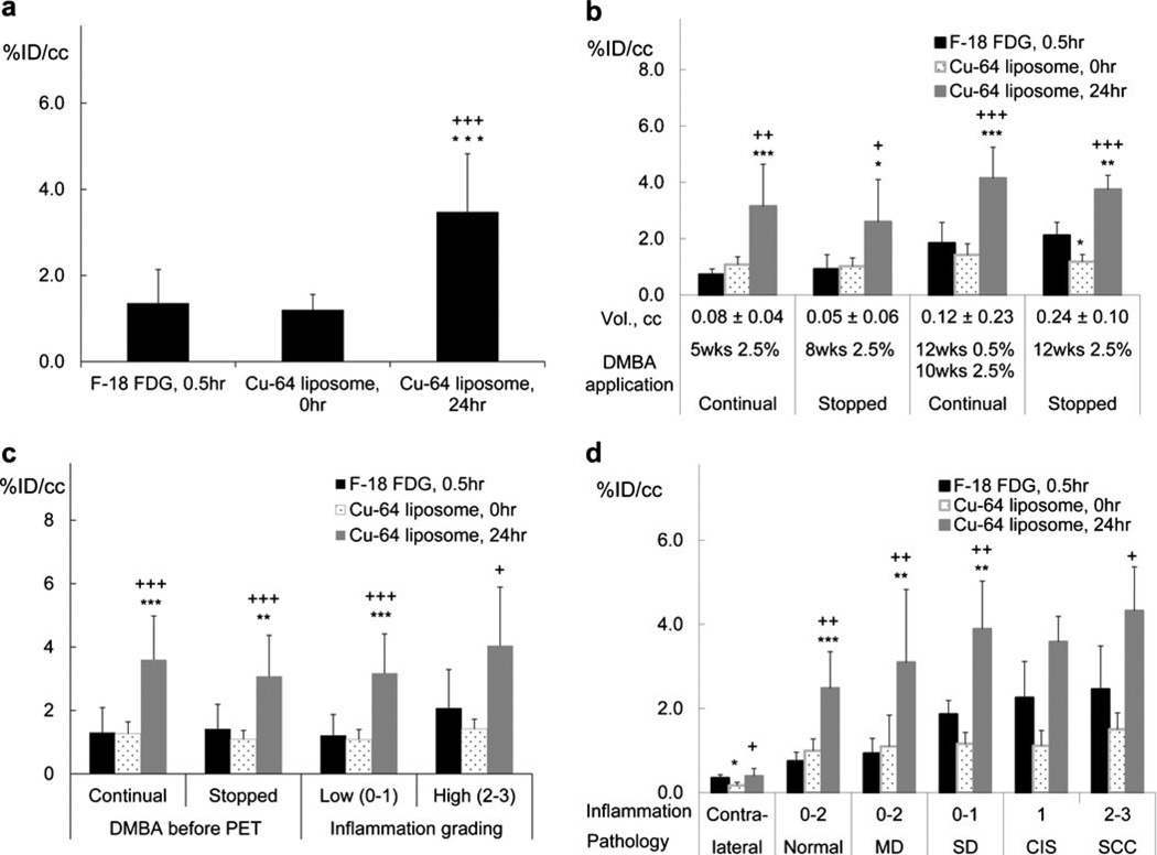
Fig. 5.
Radioactive accumulation in lesions imaged with 18F-FDG (30min) and 64Cu-liposomes (0 and 24 h): a for all animals (no grouping); b grouped by different DMBA application patterns; c grouped according to continuous or halted DMBA application; d grouped by inflammation and pathological conditions. For each case, radioactivity accumulated within the lesion was greater with 64Cu-liposomes than with 18F-FDG at the respective peak time points. Statistical significance is shown as follows: asterisk, 18F-FDG vs 64Cu-liposomes; plus sign, 64Cu-liposomes 0 vs 24 h; single symbol, p < 0.05; double symbols, p < 0.01; triple symbols, p < 0.001.

