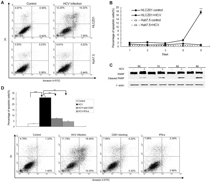
Figure 3. HCV infection triggers apoptosis of HLCZ01 cells.
(A) Annexin V expression determined by flow cytometry. HLCZ01 and Huh7.5 cells were infected with HCV at MOI of 0.1. The cells were harvested at day 9 pi and subjected to Annexin V analysis determined by Flow cytometry. The data are one representative of three independent experiments. (B) Kinetics of apoptosis in HCV-infected HLCZ01 cells. HCV-infected HLCZ01 and Huh7.5 cells were harvested for Annexin V expression determined by flow cytometry. The percentage of apoptotic cells is plotted. The data represent the means of three experiments. (C) Confirmation of HCV-induced apoptosis in HLCZ01 cells by western blot. HLCZ01 cells were infected with HCV at MOI of 0.1. Cells were collected and PARP cleavage was detected by western blot. Blots are representative of three independent experiments. (D) Blocking viral entry by anti-CD81 antibody or suppression of HCV replication by IFN reduces apoptosis of HLCZ01 cells. HLCZ01 cells were treated with anti-CD81 antibody or 100 IU/mL IFN before viral inoculation. The cells were harvested at day 9 pi for Annexin V expression determined by Flow cytometry. The graph shows the percentage of apoptotic cells, which represents the mean of 3 independent experiments.

