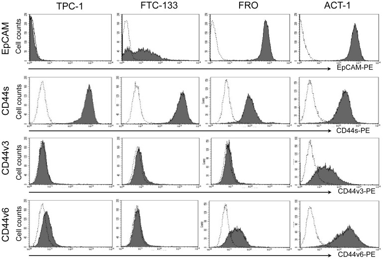
Figure 1. Expression of EpCAM, CD44s, CD44v3 and CD44v6 in thyroid cancer cell lines.
Thyroid cancer cell lines, TPC-1, FTC-133, FRO and ACT-1 were tested by flow cytometry for expression of EpCAM, CD44s, CD44v3 and v6. Filled histograms represent positive staining for the proteins of interest, open histograms show negative control with matched isotype antibody.

