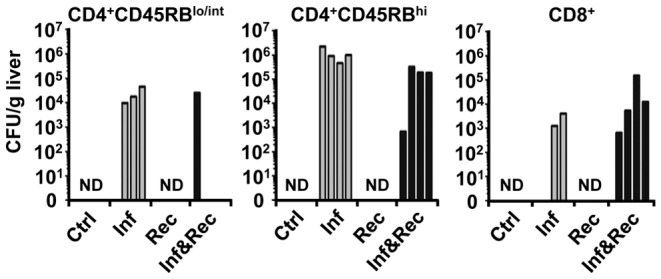
Figure 5. MAP colony forming units derived from liver tissue of Rag2−/− mice 8 weeks post infection.

Three different T cell subsets were used for adoptive transfer 4 weeks post infection: CD4+CD45RBhi, CD4+CD45RBlo/int and CD8+. Only infected as well as infected and reconstituted animals contained colony forming units i.e. MAP, as expected. Although all mice of a group were injected i.p. with 108 MAP, plating 8 weeks later did not indicate a homogeneous infection. From some infected mice viable MAP could not be revealed from liver. Similar data were obtained using PCR. Each group n = 4 mice. The experiments were carried out twice with similar results.
