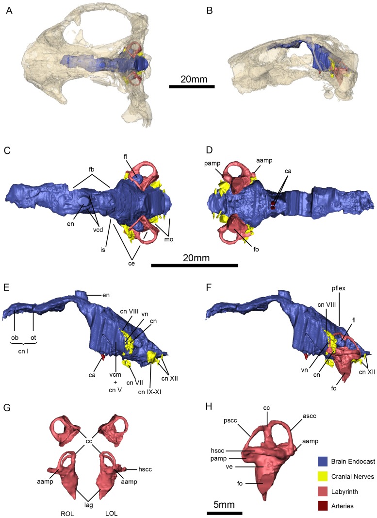
Figure 15. Niassodon mfumukasi neuroanatomy and inner ear anatomy.
Cranial endocasts in the cranium in dorsal (A), and left lateral (B) views. Cranial endocasts in dorsal (C), ventral (D), left lateral without the osseous labyrinth (E), left lateral view with the osseous labyrinth (F). Both osseous labyrinths in dorsal and anterior views (G) and both osseous labyrinths in right lateral view (H). aamp, ampulla of the anterior semicircular canal; ascc, anterior semicircular canal; ca, carotid arteries; cc, crus comunis; ce, cerebelum; cn, cochlear nerve; cn I, olfactory nerve; cn VII, facial nerve; cn VIII, vestibulocochlear nerve; cn IX-XI, glossopharyngeal and vagoaccessory nerves; cn XII, hypoglossal nerves; en, epiphyseal nerve; fb, forebrain; fl, paraflocculus; fo, fenestra oval; is, isthmus; hscc, horizontal semicircular canal; lag, lagena; LOL, left osseous labyrinth; mo, medula oblongata; ob, olfactory bulb; ot, olfactory tract; pamp, ampulla of the posterior semicircular canal; pflex, pontine flexure pscc, posterior semicircular canal; ROL, right osseous labyrinth; vcd, vena capitis dorsalis; vcm + cn V, vena capitis medialis and trigeminal nerve; ve, vestibule; vn, vestibular nerve.

