Figure 4.
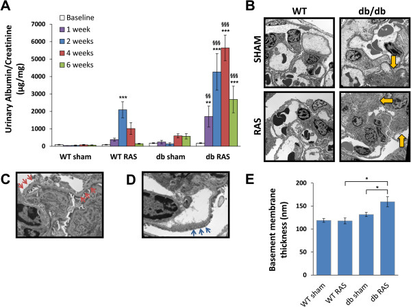
Db RAS mice developed albuminuria to a greater extent that the WT RAS mice. A. db RAS experienced persistent and progressive albuminuria compared to the transient albuminuria experienced by WT RAS. Albuminuria was measured in spot urine sample and normalized to urine creatinine. Data are presented as means ± SE. ** p <0.01, *** p < 0.001 vs. WT sham with the same time point. §§ p < 0.01, §§§ p < 0.001 vs. WT RAS or db sham with the same time point. Statistical significance was determined by 2-way ANOVA followed by Tukey adjusted post-hoc comparison. B-D. Representative electron microscope (EM) images of contralateral kidneys at 6 weeks post-surgery. db RAS mice showed significant area of mesangial matrix expansion (yellow arrows) (B) in comparison to WT RAS or db sham, along with thickened basement membrane (red arrows) (C) and podocyte fusion (blue arrows) (D). These changes were not observed in the WT mice (with or without RAS) or db sham mice. Images in B are taken at 2900× magnification while C and D are taken at 9600× magnification. E. Morphometric analysis showed significant increase in basement membrane thickness of db RAS compared to WT RAS and db sham. Data are presented as means ± SE. * p < 0.05. Statistical significance was determined by one-way ANOVA followed by Tukey adjusted post-hoc comparison.

