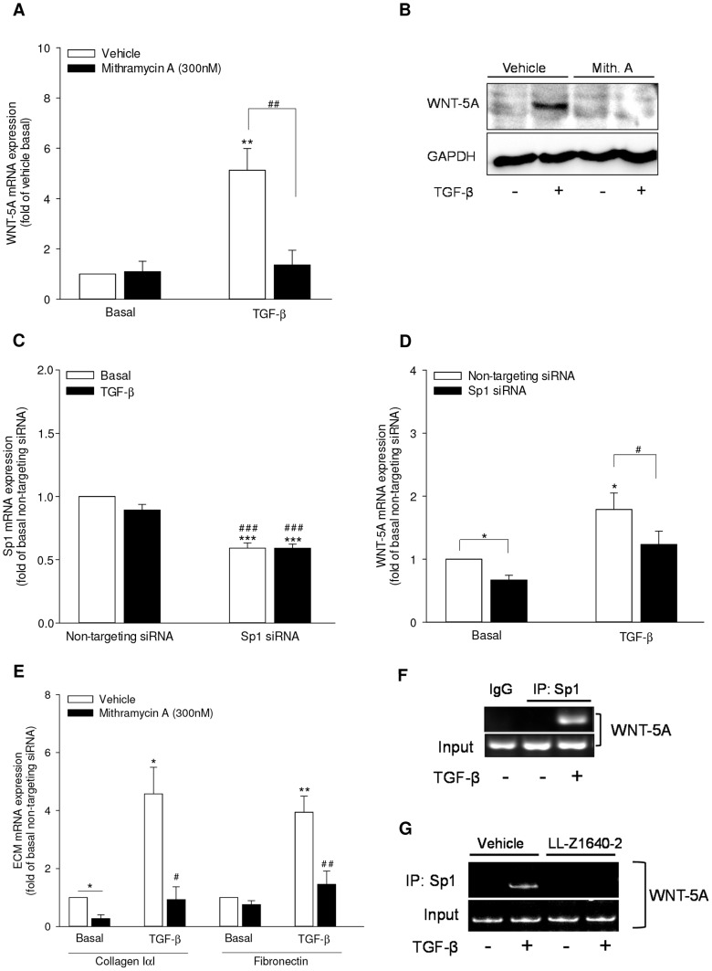
Figure 6. Sp1 is the transcription factor for TGF-β-induced WNT-5A expression in airway smooth muscle cells.
(A-B) Mithramycin A attenuates WNT-5A mRNA and protein expression. (A) Cells were stimulated with TGF-β (2 ng/ml) in the presence or absence of Mithramycin A (300 nM) for 24 hours. WNT-5A mRNA was analyzed by qRT-PCR. Data represent mean ± SEM of 4 independent experiments. **p<0.01 compared to vehicle basal, ## p<0.01 compared to TGF-β-stimulated cells; 1-way ANOVA followed by Newman-Keuls multiple comparisons test. (B) Cells were stimulated with TGF-β (2 ng/ml) in the presence or absence of Mithramycin A (300 nM) for 48 hours. Whole cell extracts were prepared and WNT-5A protein abundance was evaluated by western analysis. GAPDH was assessed as loading control. (C, D) Cells were transfected with Sp1-specific or a non-targeting siRNA as control. Subsequently, cells were stimulated with TGF-β (2 ng/ml) for 24 hours and analyzed for the expression of Sp1 mRNA (C) and WNT-5A mRNA (D) by qRT-PCR. Data represent mean ± SEM of 5 independent experiments. *p<0.05, ***p<0.001 compared to non-targeting siRNA-transfected untreated control, #p<0.05, ### p<0.001 compared to non-targeting siRNA-transfected, TGF-β-stimulated cells; 1-way ANOVA followed by Newman-Keuls multiple comparisons test. (E) Mithramycin A attenuates TGF-β-induced extracellular matrix expression. Cells were stimulated with TGF-β (2 ng/ml) in the presence or absence of Mithramycin A (300 nM) for 24 hours. Collagen IαI and fibronectin mRNA was analyzed by qRT-PCR. Data represent mean ± SEM of 4 independent experiments. *p<0.05, **p<0.01 compared to vehicle basal, #p<0.05, ## p<0.01 compared to TGF-β-stimulated cells; 1-way ANOVA followed by Newman-Keuls multiple comparisons test. (F) Sp1 is recruited to WNT-5A promoter in response to TGF-β. Cells were left untreated or stimulated with TGF-β (2 ng/ml) for 16 hours. Chromatin was prepared and ChIP analysis was performed as described in the Materials and Methods section. PCR was carried out using primers specific for Sp1 binding region on WNT-5A promoter A after immunoprecipitation with anti-Sp1 or control IgG antibody. Input DNA from chromatin preparation before immunoprecipitation was amplified to ascertain the loading. Resulting PCR products were analyzed by DNA PAGE. (G) TAK1 mediates recruitment of Sp1 to WNT-5A promoter in response to TGF-β. Cells were left untreated or stimulated with TGF-β (2 ng/ml) in the presence or absence of LL-Z1640-2 (0.5 µM) for 16 hours. ChIP analysis was performed as described above.

