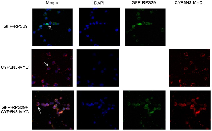
Figure 4. Intracellular co-localization of CYP6N3 and RPS29.
C6/36 cells were transfected with GFP-RPS29, CYP6N3-MYC and GFP-RPS29 + CYP6N3-MYC, repectively. Fuorescence was visualized and recorded using confocal laser scanning microscopy after 48 h of expression. CYP6N3 was localized in the cytoplasm (arrowheads) and RPS29 was distributed throughout the cell. RPS29 + CYP6N3 were co-localized in the cytoplasm (arrows). Cells nuceli were stained with DAPI.

