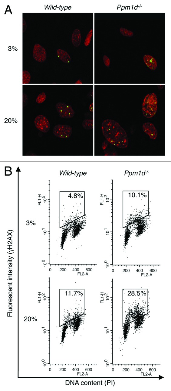
Figure 4. Increased H2AX phosphorylation associated with Ppm1d deficiency occurred predominantly in S phase. (A) Wt and Ppm1d−/− MEFs at passage 3 cultured in 3% or 20% O2 conditions were stained with anti-γH2AX antibody (Green) and PI (red). The number of γH2AX foci per cell was visually counted in at least 200 cells per group. Representative image from each sample is shown. Photographs are at the same magnification (× 400). (B) Cell cycle flow cytometric detection of γH2AX. Wt and Ppm1d−/− MEFs at passage 2 cultured in 3% or 20% O2 were stained with anti-γH2AX antibody and PI and then analyzed by dual parameter flow cytometry. The percentage of γH2AX-positive cells is indicated in the upper right of each diagram. The gate was set up to exclude background fluorescence levels by using H2AX−/− MEFs treated with the same primary antibody. The data are representative of 3 independent experiments.
