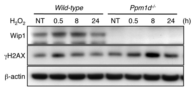
Figure 6. Prolonged H2AX phosphorylation induced by brief H2O2 exposure in Ppm1d−/− MEFs. Wt and Ppm1d−/− MEFs at passage 1 cultured in 3% O2 were treated with 100 µM H2O2 for 15 min. Cells were then washed with PBS and incubated in fresh complete medium. Cells were collected at indicated times (0.5, 8, and 24 h) after H2O2 treatment, and blotted proteins were probed with Wip1, γH2AX and β-actin antibodies. β-actin was used as loading control. NT, no treatment. The data are representative of 3 independent experiments.
