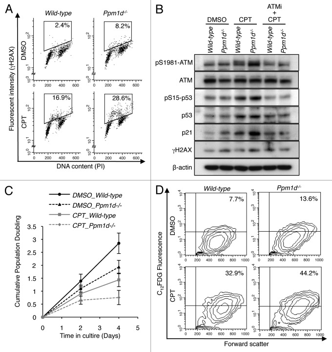
Figure 7.Ppm1d−/− MEFs showed premature senescence in the response to camptothecin-induced DNA damage. (A) Dual parameter flow cytometric detection of γH2AX and DNA content. Wt and Ppm1d−/− MEFs at passage 1 cultured in 3% O2 condition were treated with or without 20 nM camptothecin (CPT) for 2 h. As a control for CPT treatment, 0.1% DMSO was added. γH2AX intensity is represented on the y-axis, while PI staining (DNA content) is plotted along the x-axis. The percentage of γH2AX-positive cells is indicated in the upper right of each gate. The data are representative of 3 independent experiments. (B) Immunoblot analysis of ATM, p-ATM (pSer1981), p53, p-p53 (pSer15), p21, γH2AX, and β-actin in cells treated with or without 10 µM ATMi for 1 h before the addition of 20 nM CPT for 2 h. (C) Cells were cultured in 3% O2 in medium with or without 20 nM CPT and passaged at 5 × 10⁵ cells per 75 cm2 flask every 2 d. As a control for CPT treatment, medium containing 0.1% DMSO was used. Cell numbers were determined, and cumulative population doubling levels were calculated at each passage. The averages of 3 independent cultures with SD are shown. (D) Flow cytometric detection of SA-β-Gal activity in cells at passage 3, cultured as described in (C). The percentage of SA-β-Gal-positive cells is shown in the upper right of each diagram. The data are representative of 3 independent experiments.

An official website of the United States government
Here's how you know
Official websites use .gov
A
.gov website belongs to an official
government organization in the United States.
Secure .gov websites use HTTPS
A lock (
) or https:// means you've safely
connected to the .gov website. Share sensitive
information only on official, secure websites.
