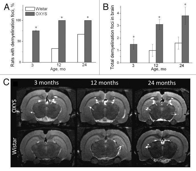
Figure 2. T2-weighed MRI images of demyelinating foci in 3-, 12-, and 24-mo-old Wistar and OXYS rats. (A) The presence of foci of demyelization in 3-, 12-, and 24-mo-old Wistar and OXYS rats (% animals). (B) OXYS rats have a greater number of demyelinating foci compared with Wistar rats. (C) Axial slices of the brain of 3-, 12-, and 24-mo-old Wistar and OXYS rats. The arrows point to foci of demyelization. The data are shown as mean ± SEM. Legend: *P < 0.05 for differences between the strains. Adapted from reference 34.

An official website of the United States government
Here's how you know
Official websites use .gov
A
.gov website belongs to an official
government organization in the United States.
Secure .gov websites use HTTPS
A lock (
) or https:// means you've safely
connected to the .gov website. Share sensitive
information only on official, secure websites.
