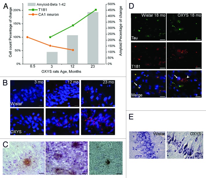
Figure 4. Time course of progression of AD-like pathology, as a percentage of either 0.5- or 3-mo-old OXYS rats in a group. (A) Rats exhibited a 47% loss of neurons in the CA1 region of the hippocampus between 0.5 and 12 mo of age. In addition, a 102% increase in the phospho-tau T181 level occurred between 3 and 23 mo of age. Total Aβ expression increased by 333% between 3 and 23 mo of age. (B) Aβ1–42 level is increased in the hippocampus of 12- and 23-mo-old but not 3-mo-old OXYS rats compared with disease-free (control) Wistar rats. Staining for Aβ1–42 (red). DAPI staining (blue) corresponds to cellular nuclei. Scale bars: 20 μm. (C) Photomicrographs demonstrate the Aβ deposits in the brain of OXYS rats detected by MOAB-2, clone 6C3. Scale bars: 50 μm. (D) Immunostaining for Tau (green) and phospho-tau T181 (red) in the cortex of 18-mo-old OXYS and Wistar rats. DAPI staining (blue) corresponds to cellular nuclei. The arrow shows colocalization of Tau and phospho-tau T181 in OXYS and Wistar rats, whereas the arrowhead shows phospho-tau T181 in OXYS rats. Scale bars: 20 μm. (E) The percentages of dead or damaged neurons in the CA1 region of the hippocampus in 15-mo-old OXYS rats are significantly greater compared with Wistar rats. Neurons were stained with cresyl violet. Adapted from reference 34 and Maksimova et al.

An official website of the United States government
Here's how you know
Official websites use .gov
A
.gov website belongs to an official
government organization in the United States.
Secure .gov websites use HTTPS
A lock (
) or https:// means you've safely
connected to the .gov website. Share sensitive
information only on official, secure websites.
