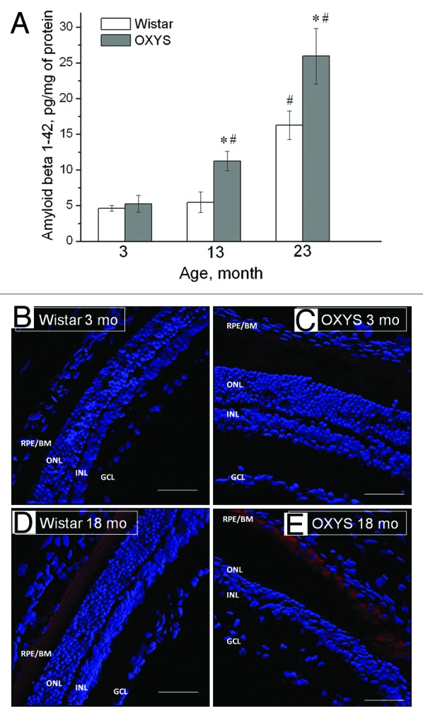
Figure 5. Aβ1–42 level is increased in the retina of OXYS rats compared with disease-free (control) Wistar rats. (A) Levels of Aβ1–42 of OXYS and Wistar rats at the age of 3, 13, and 23 mo (ELISA). *A statistically significant difference between the strains of the same age; #significant age-related differences compared with the previous age within the strain. (B–E) Confocal immunofluorescent images depict a weak Aβ (red) signal detected in a 3-mo-old Wistar rat (B), a stronger signal in a 3-mo-old OXYS rat (C), and strong staining in 18-mo-old rats of both strains (D and E). Cell nuclei are stained with DAPI (blue). The scale bar is 50 μm. RPE/BrM, retinal pigment epithelium/Bruch membrane; ONL, outer nuclear layer; INL, inner nuclear layer; GCL, ganglion cell layer. Adapted from reference 86.
