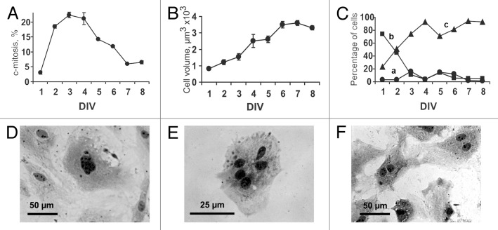
Figure 1. Eight-day in vitro (DIV) growth of myocardial cells obtained from newborn rat heart. (A) Changes in the proportion of dividing cells (c-mitoses) over 8 d of culturing. (B) Changes in mean cell volume over 8 d of culturing. Values are expressed as the mean ± SD (C) Temporal changes in the number of cultured myocardial cells with different cell volume. (a) Population of small cells with mean cell volume <250 μm3; (b) Population of cells with mean cell volume of 250–1000 μm3; (c) Population of cells with mean cell volume >1000 μm3. (D–F) Polyploid and multinucleated cardiac myocytes. Hematoxylin staining. Digital camera Leica DFC300 FX, Trinocular Microscope H 605T (WPI), objective ×40. (D) Polyploid cardiac myocyte, DIV 8. (E) Cardiac myocyte with 5 nuclei, DIV 4. (F) Binucleated cardiac myocytes, DIV 6.

An official website of the United States government
Here's how you know
Official websites use .gov
A
.gov website belongs to an official
government organization in the United States.
Secure .gov websites use HTTPS
A lock (
) or https:// means you've safely
connected to the .gov website. Share sensitive
information only on official, secure websites.
