
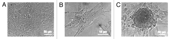
Figure 2. Three different patterns of spontaneously contracting structures in the primary culture of rat neonatal myocardial cells. (A) The confluent monolayer of contracting myocytes resulting from cell plating at a density of 5 × 10⁴ cells/cm2 (contraction rate: 28 beats/min, DIV 11). (B) The contraction of individual mature cardiomyocytes (density of cell plating: 2 × 10⁴ cells/cm2, contraction rate: 47 beats/min, DIV 3). (C) Contracting cardiomyocyte colony in the culture (density of cell plating: 2 × 10⁴ cells/cm2, contraction rate: 10 beats/min, DIV 11). Digital camera Leica DFC300 FX. The images were obtained using an inverted microscope (PIM-III, WPI), objective ×40.