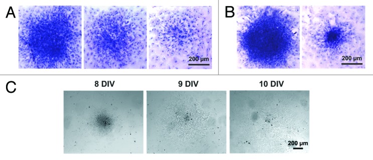
Figure 3. Structure of the cardiomyocyte colonies emerging in the primary culture of neonatal rat myocardial cells. (A) Colonies of different sizes identified on DIV 7. (B) Colonies of different sizes identified on DIV 8. Hematoxylin staining. Digital camera Leica DFC300 FX, objective ×10. (C) Dynamic changes in the size and shape of the living colony over the period of 3 DIV (DIV 8–10). Digital camera Leica DFC300 FX, objective ×10.
