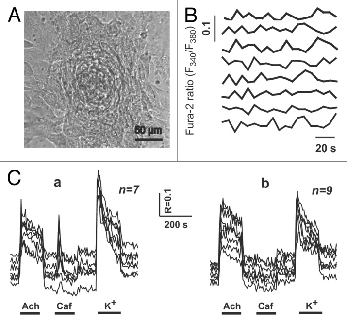
Figure 4. Spontaneous and agonist-induced calcium transients in the individual cells of the cardiomyocyte colony on DIV 10. (A) The contracting cardiomyocyte colony on the 10th day after plating of newborn rat myocardial cells. Contraction rate: 2–3 beats/min. Digital camera Leica DFC300 FX, inverted microscope PIM-III (WPI, USA), objective ×40. (B) Spontaneous [Ca2+]i fluctuations in the individual cardiomyocytes of the same colony. (C) Acetylcholine-, caffeine-, and KCl-induced (20 μM, 5 mM, and 120 mM, respectively) Ca2+ transients. (a) In the cells of the colony responsive to all applied stimuli (n = 7). (b) In the cells of the colony non-responsive to caffeine administration (n = 9).
