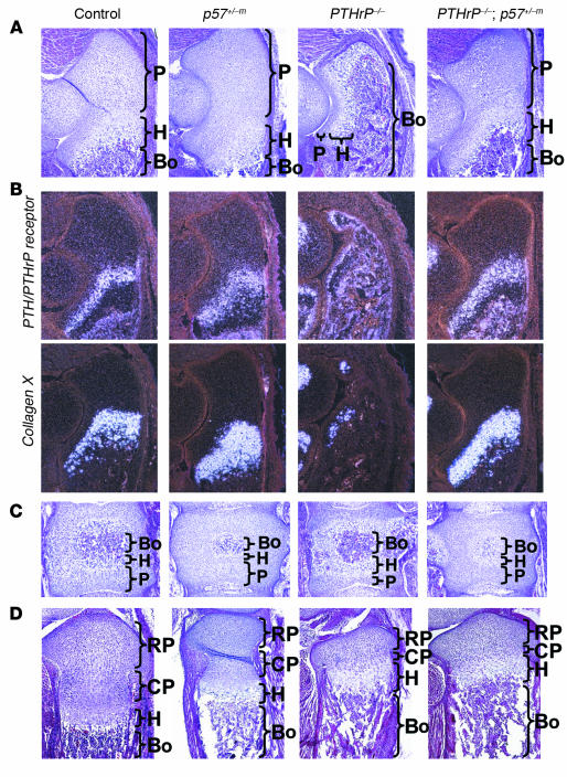
Figure 1.
Normalization of PTHrP-null morphology by removal of p57. (A_D) Sections from control, p57+/_m, PTHrP_/_, and PTHrP_/_ ; p57+/_m littermates. (A) Morphology of E18.5 ulna. The p57-null ulna has a small increase in the number of proliferative chondrocytes (P), whereas the PTHrP-null ulna has almost complete loss of proliferative chondrocytes, with expanded hypertrophic chondrocytes (H) and expanded bone formation (indicated as Bo). The PTHrP/p57-null ulna shows normal morphology. Original magnification ∞10. (B) In situ hybridization of E18.5 ulna probed with PTH/PTHrP receptor (expressed in prehypertrophic and early-hypertrophic chondrocytes) and collagen X (expressed in hypertrophic chondrocytes). The PTHrP-null ulna shows virtually complete loss of proliferative and prehypertrophic layers, and these layers are restored in the PTHrP/p57-null ulna. Original magnification, ∞10. (C) Morphology of E18.5 vertebrae. The PTHrP-null vertebra has a reduction in proliferative chondrocytes and an expanded hypertrophic zone, and the PTHrP/p57-null vertebra shows restoration of normal morphology. Original magnification, ∞20. (D) Morphology of E18.5 tibia. The PTHrP-null tibia has a large reduction in the zone of columnar proliferative chondrocytes (CP), which is not normalized in the PTHrP/p57-null tibia. RP, round proliferative chondrocytes. Original magnification, ∞10.

