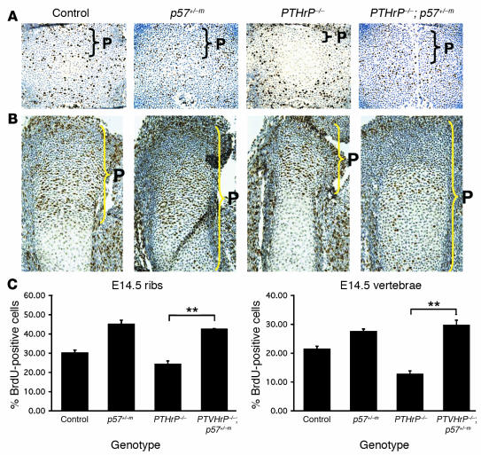
Figure 2.
Reversal of early exit of proliferation, proliferation rate in PTHrP/p57-null mutants. (A and B) Sections from control, p57+/_m, PTHrP_/_, and PTHrP_/_; p57+/_m littermates. (A) BrdU incorporation in E15.5 vertebrae, with immunohistochemical detection of BrdU+ cells (brown), and all cells counterstained with hematoxylin (blue). The PTHrP-null vertebra has early exit of proliferation, indicated by a reduction in the number of proliferating layers of chondrocytes, which is increased in the PTHrP/p57-null vertebra. Original magnification ∞20. (B) BrdU incorporation in E14.5 tibia. There is early exit of proliferation in the PTHrP-null tibia, as determined by the distance from the articular region at which cells stop incorporating BrdU. The PTHrP/p57-null tibia has reversal of the early exit of proliferation, with a longer region of BrdU-positive chondrocytes. The p57-null tibia also has a delay in cell cycle exit. Original magnification, ∞10. (C) Rate of chondrocyte proliferation in E14.5 ribs and vertebrae. BrdU incorporation rate was quantitated as described in Methods. The PTHrP-null ribs and vertebrae have a significant reduction in proliferation rate, which is elevated in the PTHrP/p57 -null bone tissue. Bars represent mean ± SEM. **P < 0.001.

