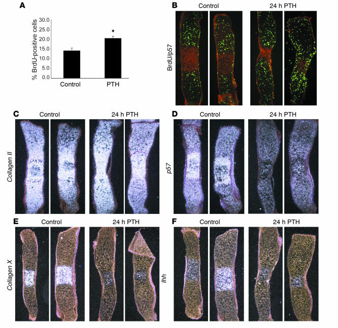
Figure 5.
Metatarsal culture response to PTH treatment. (A) Chondrocyte proliferation rate in metatarsal cultures. E15.5 wild-type metatarsals were cultured 48 hours, then treated 24 hours with vehicle or 10_7 M PTH(1–34). Bars represent mean ± SEM; *P < 0.05. PTH treatment elevates chondrocyte proliferation rate (percentage of BrdU-positive cells). (B_F) Sections from metatarsals treated with vehicle or 10_7 M PTH(1–34) for 24 hours. (B) BrdU/p57 double-immunofluorescent detection. PTH treatment increases the size of the proliferative pool, as indicated by increased length of BrdU-positive (green) region; and delays cell cycle exit, as indicated by the reduction in the region of postproliferative chondrocytes, which express p57 protein (red) and do not incorporate BrdU. (C) In situ hybridization showing collagen II mRNA expression. There is no change in collagen II expression in the proliferative chondrocytes of metatarsals treated with PTH. (D) In situ hybridization showing p57 mRNA expression. PTH treatment decreases p57 expression in both the proliferative chondrocytes and the prehypertrophic and early-hypertrophic chondrocytes. (E) In situ hybridization showing collagen X mRNA expression. PTH treatment reduces the area of hypertrophic, collagen X_expressing chondrocytes and also reduces the intensity of collagen X expression. This indicates that PTH delays cell cycle exit and suppresses hypertrophic differentiation. (F) In situ hybridization showing Ihh mRNA expression. PTH treatment decreases Ihh expression in the prehypertrophic and early-hypertrophic chondrocytes, indicating a general suppression of hypertrophic differentiation. Original magnification, ∞20 in B_F.

