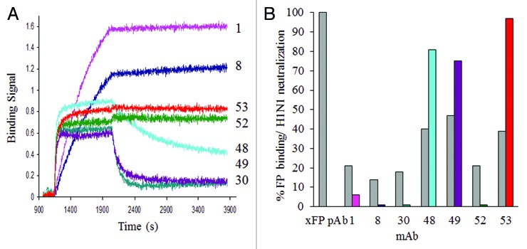
Figure 2. Kinetic binding and neutralizing activity of a panel of antibodies cross-reactive to the fusion region of group 1 HA. (A) Comparison of the binding kinetics of mAb 1, 8, 30, 48, 49, 52 or 53 (captured on an anti-human Fc sensors), to 200nM of HA monomer of the H1N1 A/CA/04/2009 strain. (B) Comparative fusion peptide binding (gray bars) as a percent relative to a rabbit pAb directed to and purified by the fusion peptide; and neutralizing activity of antibodies (colored bars), at a constant IgG concentration of 5 µg/ml in MDCK cells infected with the H1N1 A/CA/04/2009 virus. Plaques were counted in duplicate wells and expressed as a percent of plaques in an uninhibited infection. A comparison of the breadth and potency of HA coverage for selected neutralizing group 1 antibodies can be found in Table 4 and Figure S2.

An official website of the United States government
Here's how you know
Official websites use .gov
A
.gov website belongs to an official
government organization in the United States.
Secure .gov websites use HTTPS
A lock (
) or https:// means you've safely
connected to the .gov website. Share sensitive
information only on official, secure websites.
