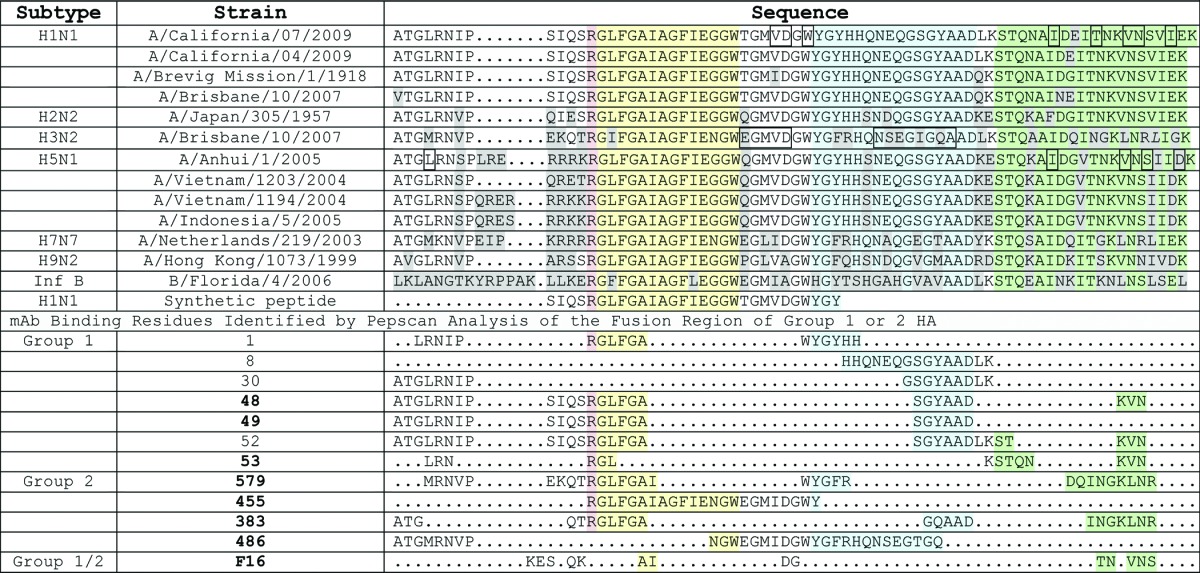
Table 1. Alignment of HA protein surrounding stalk region and epitopes of antibodies.

Residues are highlighted according to conserved HA structural domains (see Fig. 1) surrounding the stalk domain(in green). The Arginine colored red is conserved among HA proteins and is cleaved by extracellular proteases to trigger the fusion conformation. In sequence order: a loop region (yellow)is next to an anti-parallel β-sheet (blue),followed by the alphα-helical stalk region (green). Anti-HA antibodies (neutralizers in bold) with Pepscan analysis data are shown in alignment. Boxed residues are key contacts in the F10 antibody (H1N1), CR6261 (H5N1) or CR8020 (H3N2).
