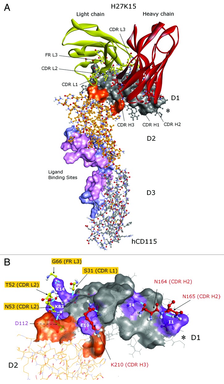
Figure 3. Predicted epitope of the H27K15 mAb. Best docked pose of the H27K15 4m model on the hCD115 crystal. (A) Red, heavy chain; yellow, light chain of H27K15 Fv; gray, D1 domain; orange, D2 domain of the hCD115. Surface corresponds to the interface area between H27K15 and hCD115. Residues specifically interacting with IL-34 (light blue) and interacting with both IL-34 and CSF-1 (light pink) are also indicated as ligand binding sites surface. (B) hCD115 residues forming hydrogen bonds are indicated in purple. *indicates the position of the G-D-P-L unresolved loop. Views were generated using Discovery Studio v3.5 (Accelrys).

An official website of the United States government
Here's how you know
Official websites use .gov
A
.gov website belongs to an official
government organization in the United States.
Secure .gov websites use HTTPS
A lock (
) or https:// means you've safely
connected to the .gov website. Share sensitive
information only on official, secure websites.
