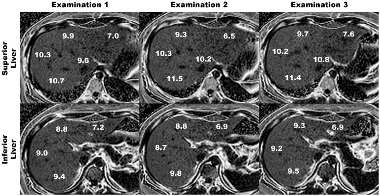
Figure 2.
Six-echo parametric PDFF maps through superior (upper panel) and inferior (bottom panel) parts of the liver obtained during three separate examinations in a single day. Upper panel shows PDFF estimates in segments 1, 2, 4a, 7, and 8. Lower panel shows PDFF estimates in liver segments 3, 4b, 5, and 6. Notice close agreement between segmental PDFF estimates across examinations.

