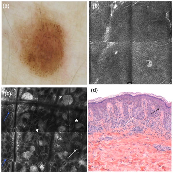
Figure 3.

(a) Dermoscopy showing irregular globular pattern (b) RCM mosaic 1000×1000 μm showing a preserved honeycomb pattern in epidermis (*). (c) RCM mosaic at dermoepidermal junction showing irregular nests (*), with irregular edged papillae (blue arrows), junctional thickenings (▲) and some atypical hyperreflective cells within the dermal papillae (white arrows). (d) Histopathology (haematoxylin and eosin 20X) showing atypical cells (black arrow) and nests (white arrow) from a in situ melanoma.
