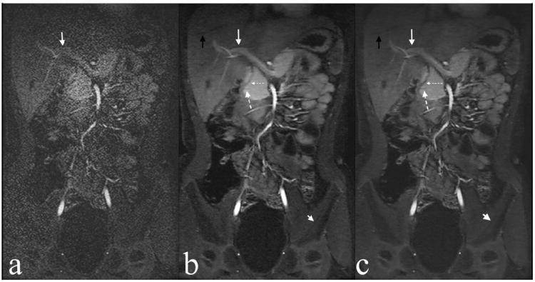
Figure 1.

Example of ARC (a), L1-SPIRiT (b), and CC-L1-SPIRiT (c) reconstructions of a 6-year-old female. Note delineation of hepatic vein branch (black arrow), portal vein (white arrow), pancreatic duct (large dashed arrow), bile duct (small dashed arrow), and cortex of left iliac bone (short white arrow).
