
Figure 2.
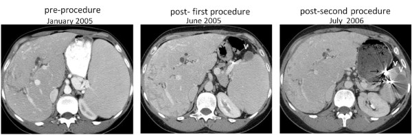
Patient 2, serial axial CT scans of the abdomen, demonstrating a heterogeneous liver, radiopaque material placed in the branches of the splenic artery (→), which has resulted in wedge shaped splenic infarctions (v). The splenomegaly is seen to progressively resolve, and there is progressively less impingement upon the left kidney.