Figure 3.

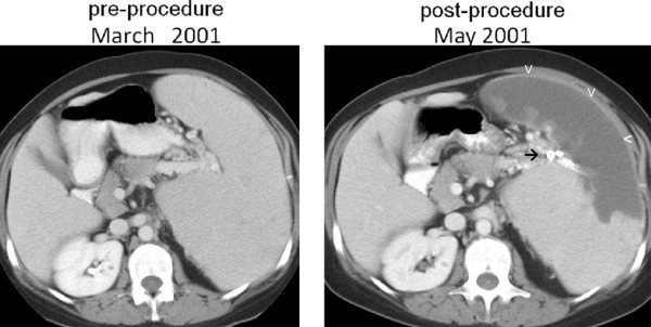
Patient 3, pre and post-procedure axial CT scans with contrast, demonstrating massive splenomegaly, the successful placement of radiopaque material in branches of the splenic artery (→), resulting in splenic infarction (v).

Patient 3, pre and post-procedure axial CT scans with contrast, demonstrating massive splenomegaly, the successful placement of radiopaque material in branches of the splenic artery (→), resulting in splenic infarction (v).