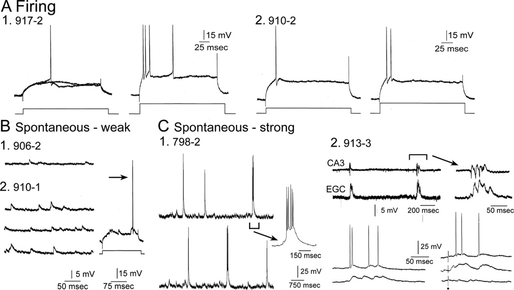
Figure 6.
Comparative physiology of hilar EGCs. A: Representative examples of directly-evoked APs are shown for two different hilar EGCs (1,2). To evaluate spike frequency adaptation, higher currents were injected as previously described (Scharfman et al., 2000). For each cell the AP evoked at threshold is on the left and the response to increased current injection is on the right. B: Spontaneous activity is shown from two hilar EGCs to illustrate examples where hilar EGC spontaneous activity was relatively weak. Even though activity was subthreshold at resting potential, depolarization of the cell led to suprathreshold activity (arrow). C: Spontaneous activity is shown from hilar EGCs that is robust, i.e., repetitive spontaneous burst discharges. 1) A continuous recording from a hilar EGC (798-2) exhibits periodic spontaneous burst of APS. The arrow points to one of the bursts with a different temporal calibration (100 msec) to show it in more detail. 2) An extracellular recording from the CA3b pyramidal cell layer is shown (CA3; top trace) simultaneous to a recording from a hilar EGC (lower trace; expanded on the right) to demonstrate the synchrony between the hilar EGC and CA3. Lower left: A hilar EGC with repetitive burst discharges was recorded at two membrane potentials. Two spontaneous bursts of activity are shown, one at a depolarized potential where APs occurred during the burst, and one at a hyperpolarized potential where APs did not occur during the burst. Lower right: Bursts were recorded in response to stimulation of the outer molecular layer to show that they could be evoked by activation of the perforant path, as previously described (Scharfman et al., 2003). In addition, the lower right traces show that the bursts were all-or-none, because some stimuli that failed to trigger a response did not show a graded decline in amplitude, but simply failed altogether. All-or-none responses suggest that paroxysmal depolarization shifts were responsible for the bursts, as previously discussed (Scharfman et al., 2000, 2007; Scharfman, 2004).

