
Figure 4.
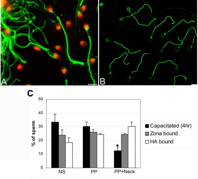
Comparison of tyrosine phosphorylation (TP) pattern in zona pellucida-bound (A) and HA-bound (B) sperm. Fluorescence was localized to the principal piece and neck region of spermatozoa in an identical pattern within both the zona pellucida- and the HA-bound sperm fractions. (C) Comparison of the extent of TP within the whole sperm population (motile and nonmotile) of capacitated sperm (4 hours), zona pellucida-bound and HA-bound sperm fractions. NS indicates nonstaining; PP, principal piece; PP + neck, principal piece and neck region. Data represent the mean proportion of each pattern within the 16 men. Two hundred sperm were examined for each patient (3200 sperm in all). Asterisk indicates significant differences (P < .05). No significant differences were found between the proportions of sperm with TP within the HA- and zona pellucid-bound sperm fractions.