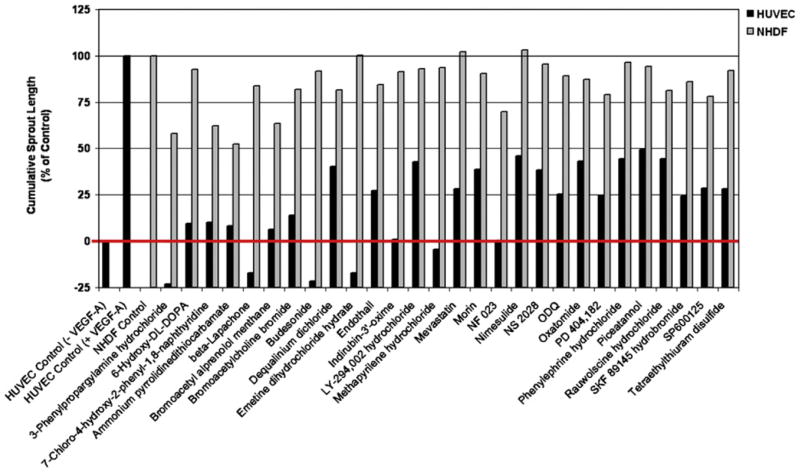
Figure 3. Twenty-Eight Compounds Inhibit VEGF-A-Induced HUVEC Sprouting Angiogenesis.
VEGF-A-driven HUVEC angiogenesis was quantified as the average cumulative sprout length (black bars). The VEGF-A-driven sprouting was set to 100%, and the basal sprouting in the absence of VEGF-A was set to 0%. The red line is a reference line for the basal HUVEC sprouting. Scattering of clustered NHDFs in collagen gels in the presence of basal medium was set to 100% (gray bars). Twenty-eight compounds from the LOPAC library inhibited sprouting at > 50% (black bars) and NHDF scattering at < 50% (gray bars).

