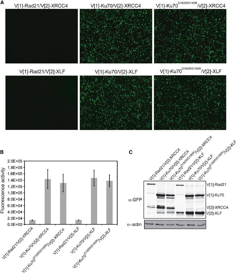
Figure 4. Ku70 α5 Is Not Required for Ku’s Association with XRCC4 or XLF.
(A) Visualization of PCA using Ku70 or Ku70D192R/D195R with XLF or XRCC4. Images are at 4× magnification.
(B) Fluorescence quantification of PCA shown in (A). The error bars represent SD of the mean.
(C) Protein levels resulting from transfections in (A) detected by immunoblot analysis of WCE using a GFP antibody. β-actin was used as a loading control.

